- Location - Engineering East Room A009
- Imaging Resolution - 0.9nm (15kV)
- Magnification - x25 to 1,000,000
- Acc. Voltage - 0.01kV to 30kV
- FIB Resolution - 3nm (statistical method)
- Single GIS: Pt and C
Learn more about the Zeiss Crossbeam 550 FIB-SEM
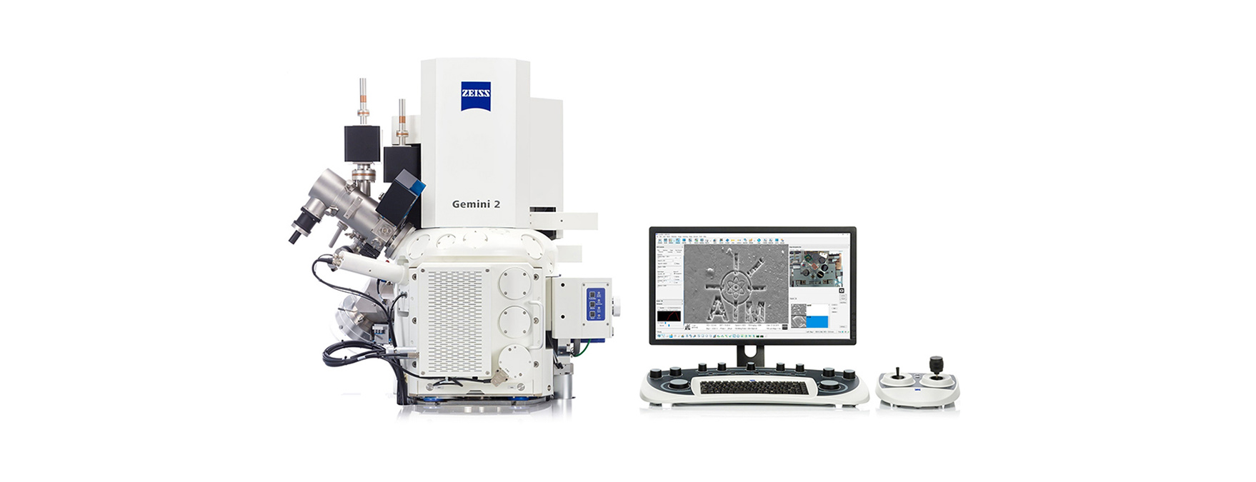
ZEISS FIB-SEMs, Focused Ion Beam Scanning Electron Microscopes, combine 3D imaging and analysis performance of the Gemini electron beam column with the ability of a FIB for material processing and sample preparation on a nanoscopic scale. ZEISS FIB-SEMs for nanotomography and nanofabrication.
What is a Focussed Ion Beam? FIB-SEMs combine the imaging capabilities of an SEM with a second co-aligned column to accelerate ions to selectively mill out areas of a sample. It is used particularly in the semiconductor industry, materials science and increasingly in the biological field for site-specific analysis, deposition, and ablation of materials.
Crossbeam features for lamella preparation:
- A focused ion beam with a 100 nA milling current
- An automated workflow that prepares large amounts of TEM lamellas on user-defined positions of your sample.
- Prepare stable lamellas to electron transparency with low energies to control amorphous layer.
The Crossbeam is also part of Zeiss' correlative workflow which spans from XRM to light microscopy to electron microscopy. Zeiss' 'Shuttle & Find' software allows for the seamless relocation of identified regions of interest between multiple scale ranges to best take advantage of the advantages of each instrument. While integrating this with the 'Connect' add-on for Zen Core, it allows for the data to be visualised in a much more informative manner with several images from different scale-lengths/techniques allowed to be placed over each other. In the Crossbeam this correlative process is enhanced further by the use of ATLAS software for an enhanced serial sectioning workflow that increases the precision of the slice data capture and image quality.
Image Courtesy of Zeiss